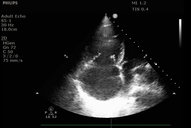
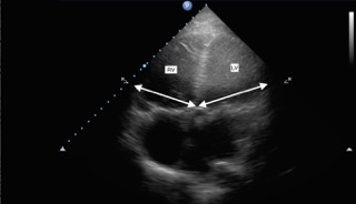
Rv Lv Ratio Echocardiography
Https Encrypted Tbn0 Gstatic Com Images Q Tbn 3aand9gctrtldus7mggg4dy8o1fwklwf3yeeeu 8qcng Usqp Cau
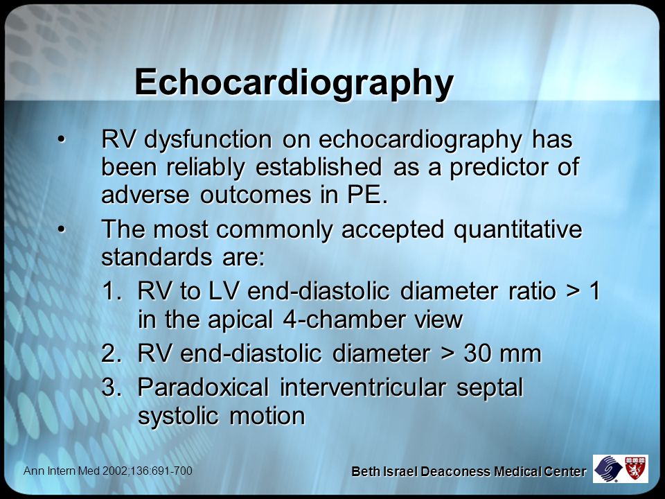
Basic Haemodynamic Assessment With Echo Iheartscan
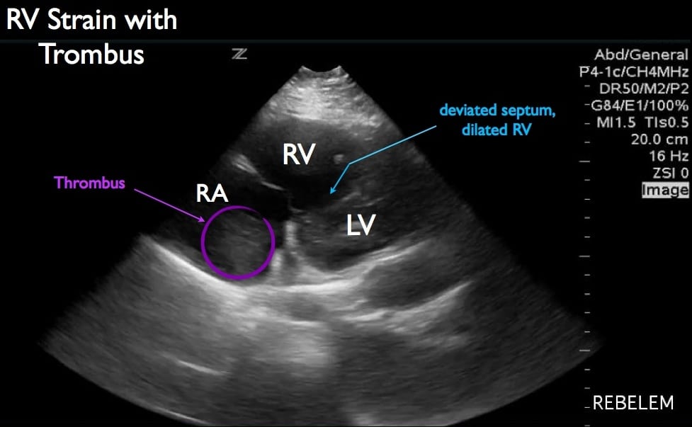
Diagnosis Of Right Ventricular Strain With Transthoracic Echocardiography Rebel Em Emergency Medicine Blog

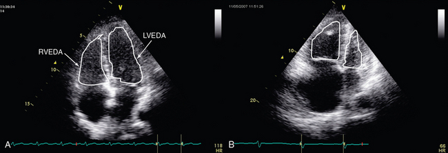
Figure 1 From Increased Right To Left Ventricle Diameter Ratio Is A Strong Predictor Of Right Ventricular Failure After Left Ventricular Assist Device Semantic Scholar

Imaging Right Left Ventricular Interactions Jacc Cardiovascular Imaging

Acep American College Of Emergency Physicians
While measurements are typically made using a true 4 chamber reformat made from the thin slice ct data.
Rv lv ratio echocardiography.

Ctpa Demonstrating The Rv Lv Ratio Measurement Note Ctpa Computed Download Scientific Diagram
Differentiating Acute Versus Chronic Right Heart Failure With Bedside Echocardiography Emra
Submassive Pe Emory School Of Medicine
Evaluation Of Right Ventricular Function In The Intensive Care Unit By Echocardiography Consultant Level Examination Radiology Key
Https Encrypted Tbn0 Gstatic Com Images Q Tbn 3aand9gcryyxpe I0f Apg9ghlpraeedhflrw1vi16uq Usqp Cau

Graphic Representation Of Transthoracic Echocardiographic Parameters In The Assessment Of Right Ventricular
Https Www Ahajournals Org Doi Pdf 10 1161 01 Cir 68 6 1201

Allied Health Allied Health Testing Diagnostics Echocardiogram

Pdf Echocardiography In Pediatric Pulmonary Hypertension
3 3 2 Right Ventricular Size 123sonography
Diastolic Dysfunction Point Of Care Ultrasound Radiology Reference Article Radiopaedia Org

Assessment Of Rv Structure And Function By Echo At 4 Weeks Post Su A Download Scientific Diagram

Correlation Of A New Echocardiographic Index Of Right Ventricular Remodeling In Pulmonary Hypertension With Invasive Hemodynamics In Rat Model Jacc Journal Of The American College Of Cardiology
Echocardiography In Pulmonary Embolism Ppt Video Online Download

Apical 5 Chamber Cardiac Sonography Diagnostic Medical Sonography Echocardiogram

Figure 1 From Acute Right Ventricular Dysfunction Real Time Management With Echocardiography Semantic Scholar

Right Ventricular Involvement In Hypertrophic Cardiomyopathy Patterns And Implications Sciencedirect
Https Journal Chestnet Org Article S0012 3692 19 31374 1 Pdf
1

A Rv Lv S Ratio In Control And Pab Mice B Echocardiography Of Download Scientific Diagram

Abstract 14015 Right Ventricular Remodeling Is Additive To Global Longitudinal Strain In Predicting Long Term Survival In Patients With Pulmonary Hypertension Circulation

Qp Qs Ratio In Echo Echocardiography Barnard Health Care Echocardiogram Diagnostic Medical Sonography Cardiac Sonography
Https Criticalcarecanada Com Presentations 2016 Peep And The Rv During Mechanical Ventilation Pdf
Https Onlinelibrary Wiley Com Doi Pdf 10 1111 Pan 13641
Source : pinterest.com
